What Tests Detect Pericardial Effusion
The basic test to determine the presence of a pericardial effusion is echocardiography or echocardiogram. Other tests are the electrocardiogram, chest X-ray, CT scan, and MRI.
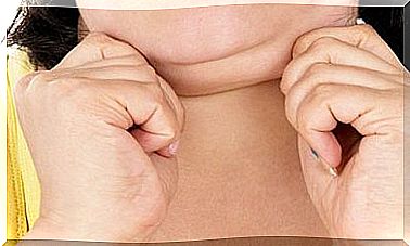
Pericardial effusion is a condition in which there is an excessive accumulation of fluid between the heart and the pericardium. The latter is the sac that covers the heart. It is estimated that there is leakage when the liquid exceeds 50 ml.
Between the heart and the pericardium there is always a thin layer of fluid. However, when there is an illness or injury, inflammation occurs. This, in turn, causes the amount of fluid to increase. Despite this, sometimes there is a pericardial effusion without previous inflammation.
Pericardial effusion puts pressure on the heart and affects its function. If left untreated, it could cause heart failure and lead to death. The tests that allow to detect this condition are the following.
Pathways to detect pericardial effusion

Echocardiogram
Echocardiography , echocardiography or cardiac ultrasound, is the preferred test to detect a pericardial effusion. The National Library of Medicine of the United States explains that it is a test that allows you to visualize the structure of the heart and study its ability to pump blood.
The echocardiogram with the Doppler technique also makes it possible to establish the exact velocity of the heart’s flows. From a technical point of view, the two-dimensional M-mode echocardiogram is the ideal technique for diagnosing, quantifying, and monitoring pericardial effusion.
The absence of echoes between the epicardium and the lateral pericardium is a finding that allows the diagnosis of a pericardial effusion. The cardiologist determines the size of the effusion from the amount of space between the two layers of the pericardium.
It should be noted that there are basically two types of echocardiogram : the transthoracic, in which a device is used that is placed on the chest, at the level of the heart, and emits sound; and the transesophageal, in which the device is introduced into the digestive tract into the esophagus. The latter provides more detailed data.
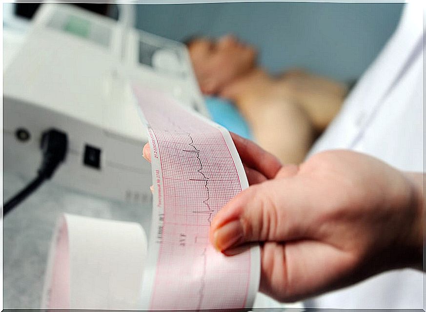
Electrocardiogram
The electrocardiogram records the electrical activity of the heart. Basically, according to data from the medical encyclopedia cited above, it allows evaluating heart rhythm and function. The pericardial effusion causes alterations in the graph, but these are not specific.
Typically, certain abnormalities are seen in the QRS complex. This is a vector that reflects the sum of all the electrical discharges that occur in the cells of the ventricles. In general, when there is a pericardial effusion, a reduction in QRS voltage is seen .
Likewise, in these cases a flattening of the T waves is observed. If the effusion is very severe and there is plugging, an electrical alternation is observed. Typically, the P wave appears wide and bimodal, suggesting an abnormality.
Chest X-ray, CT and MRI
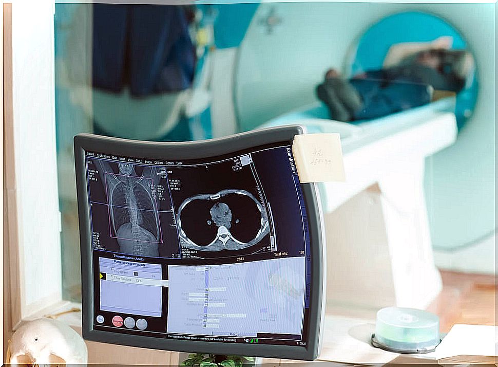
The National Heart, Lung and Blood Institute notes that chest radiography is an x-ray imaging that allows all organs in the thoracic area to be visualized. Computerized axial tomography or CT is a test that also uses X-rays and that allows images to be obtained in the form of cross sections or in three dimensions.
For its part, magnetic resonance imaging (MRI) is an examination by which detailed images of the interior of the body are obtained, in two or three dimensions. It provides much more specific information than radiography or CT.
When there is pericardial effusion, there is an alteration in the cardiac silhouette that one or all of these tests can detect. Silhouette enlargement usually occurs when more than 250 ml of fluid accumulates in the pericardial sac.
However, only with more than 50 ml can we speak of a pericardial effusion. Therefore, a chest x-ray, for example, does not show the enlarged silhouette, even though the abnormality is present. The TAC and the MRI are much more conclusive.
Quantification of pericardial effusion
There are no universally accepted criteria for quantifying the volume of pericardial effusion. This is because all methods have limitations in determining the actual amount of fluid in the pericardial sac.
The most accepted technique for quantification is that proposed by Weitzman. It is based on M-mode echocardiography. It proposes adding the echo free spaces in the anterior and posterior sacs at the end of diastole. We speak of a slight effusion when the sum is 10 mm or less; moderate, between 10 and 19 mm and severe, when it is 20 mm or more.
It is important to rule out the presence of cardiac tumors and pericardial cysts before making the diagnosis. In addition, it should be verified that there is no pleural effusion or presence of epicardial fat.